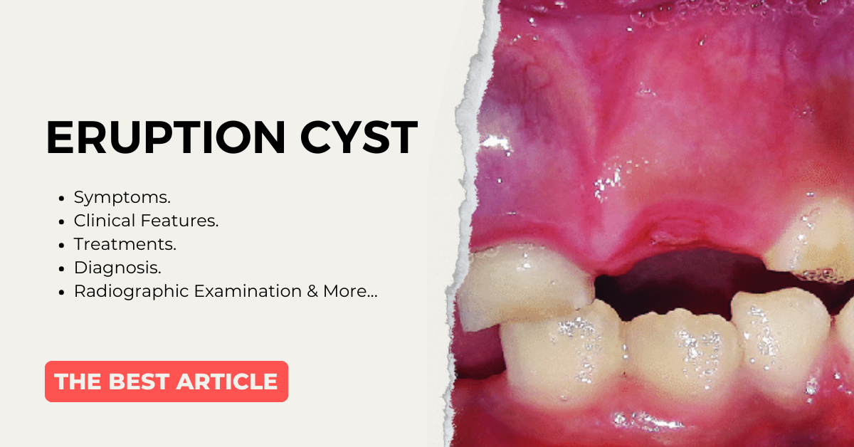
Did you ever get tensed by seeing a bluish discolored swelling in the gums of your child? Here is the solution for you. Pedodontics can help to treat this lesion.
In this article, I will discuss a case report of an 8-year-old boy who came to my clinic with an eruption cyst.
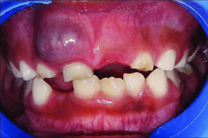
At around 10 O’clock a boy who is 8 years old with his parents came to my clinic. The parents were so tense. I made the boy sit in my dental chair and asked for the chief complaint. The parents said, “Madam there is bluish discolored swelling seen in the front tooth region. This was initially small but recently it got increased in size”. Then I asked about the extraction of 51. They said their 51 got extracted one year back due to caries. And after a few months, this discoloration started appearing. Initially, we didn’t take it into consideration because it was small, but later after a few months, we got tensed as its size increased. The parents were very much tensed and they asked me whether it was a tumor or something.
When I checked for an oral examination, there was no pus discharge and the physical body examination was normal. Also, I asked about any trauma or allergic to any spicy food, etc. and there is no such history.
Now the child is in mixed dentition and all the permanent 1st molars are erupted except the 11 [the upper right permanent central incisor].
When I observed the soft tissue there was no abnormality except in the region of 51. The size of the swelling is approximately 1.5x 1.5cm. when I examined with my finger by wearing a glove it was soft and fluctuant. After the clinical examination, I asked the parents to go for a radiograph. The radiographic examination revealed there is no underlying bony involvement. Then I came to know that it was nothing but an Eruption cyst.
Then I asked the parents to wait for two weeks and said to them, would see if it ruptured on its own then there was no need to go for any surgical intervention. But they came to me after 20 days and said there was no change. Finally, we decided to go for surgical intervention to permit the root to erupt. First, the medical history of the patient is taken and there is no abnormality detected. A blood examination is done. Local anesthesia is given. Waited for some time to act the anesthesia. A BP blade is taken and a small incision is made on the cyst, draining the content of the cyst. In the next step, a window is created for the exposure or eruption of 11.
Post-operative instruction and medications are given. The patient is recalled after 14 days.
So, after reading this I hope you might get some idea about the eruption cyst. Now let me discuss in detail about this.
Contents
WHAT DO YOU MEAN BY ERUPTION CYST?
From the term will come to know that it’s a cyst which is classified under dentigerous cyst. They are frequently associated with the eruption of permanent or deciduous teeth in children.
From the above statement don’t get confused between eruption cyst and dentigerous cyst.
An eruption cyst is in fact a dentigerous cyst occurring when a tooth is impeded in its eruption within the soft tissues overlying the bone. Whereas the dentigerous cyst develops around the crown of an unerupted tooth that is lying in the bone.
This cyst is often called an eruption hematoma.
Now you all will be eager to know how it happens. So now will discuss the pathogenesis of eruption cysts.
WHAT IS THE PATHOGENISIS OF AN ERUPTION CYST?
The eruption cyst occurs due to dilation of the tooth follicle. That is, it occurs due to the dilation of normal tooth follicles caused by to accumulation of tissue fluid or blood.
WHERE DO YOU SEE COMMONLY?
In 30 percent of cases, they are seen during the eruption of molars and canines, and in 11 percent of cases they are seen during the eruption of central incisors.
WHAT ARE THE CLINICAL FEATURES?
Appearance
First will talk about how it appears. When we observe clinically the lesion appears as a translucent swelling seen on the alveolar ridge at the site of eruption.
Age
They are commonly seen in the age group below 10 years of age.
WHAT IS ERUPTION HEMATOMA?
In some cases, the cystic cavity contains blood and in this case, the swelling appears purple or deep blue in color, so it’s named an eruption hematoma.
As we discussed above, they are commonly seen in the molar, canine, and incisor regions.
WHAT ARE THE RADIOGRAPHIC EXAMINATION?
The radiographic examination shows a saucer-shaped excavation of bone projecting near the cavity. And this is seen only in very few cases.
When we observe the margin it’s well defined.
There is an expansion of the normal follicular space of erupting tooth crowns.
HOW WILL YOU DIAGNOSE?
When we diagnose clinically there is a soft fluctuant translucent swelling around the molar, canine, or incisor.
When we observe radiographically in some cases, we can see a saucer-shaped excavation of bone with a well-defined margin.
Treatments of Eruption Cyst
The treatment includes surgical intervention for the teeth to erupt, as we discussed above. Before going to surgical intervention, we will wait for the cyst to rupture spontaneously to facilitate the tooth eruption.
CONCLUSION
As a part of the conclusion, I conclude that the eruption cyst is seen commonly in deciduous or permanent teeth. Its fluctuant swelling is seen in the alveolar ridge. Radiographically shows a saucer-shaped excavation in some cases.
If you are a regular reader then you must know about Dentigerous Cyst also. I have written a detailed blog about the same.
If you have any questions related to this article, then you can comment your questions below, and I will revert back as soon as possible.